
Gram Positive Bacteria & Gram Negative Bacteria
Gram Positive and Gram Negative Bacteria
Table of Contents
· Bacteria are minute, single celled, microscopic organisms, which are classified as prokaryotic cells. These organisms comprise a simple physical structure, including cell wall, capsule, DNA, pili, flagellum, cytoplasm and ribosomes and lacks true nucleus.
· Depending upon the staining technique, Bacteria can be gram-positive or gram-negative. Let us have a detailed look at the difference between the two types of bacteria.

Cell Wall of Bacterial Cell
· Most of the Prokaryote have a rigid cell wall that protects the bacterial protoplasm from any damage.
· The major component of the bacterial cell wall is Peptidoglycan or Murein (made of mucopolysaccharide + polypeptide).
· The peptidoglycan gives the rigidity and strength to the Bacterial cell wall, thus, describing its shape and protecting the protoplasmic content.
· It is a polysaccharide made of two glucose derivatives, N-acetyl glucosamine (NAG) and N-acetylmuramic acid (NAM), alternated in long chains. The chains are cross-linked to one another by a tetra-peptide that extends off the NAM, allowing a lattice-like structure to form.
· The four amino acids of this tetrapeptide are D-alanine, L-alanine, D-Glutamic acid and L-lysine (in Gram positive bacteria) or diaminopimelic acid (in Gram negative bacteria).
· The NAG and NAM strands are synthesized in the cytosol of the bacteria. They are connected by inter-peptide bridges.
· From the peptidoglycan inwards all bacterial cells are very similar.
Gram Staining Technique
·Gram Staining technique was proposed by Christian Gram, a microbiologist, using Gram stain to distinguish the two types of bacteria based on the difference in their cell wall structures.
Objective
· This gram staining method differentiates the bacteria into Gram Positive and Gram Negative Bacteria, which helps in the classification and differentiation of microorganisms. The Gram stain separates bacteria into two groups –
· Gram positive Bacteria that retain the primary dye (Crystal violet) and
· Gram negative Bacteria that take the color of the counterstain (usually Safranin).
Principle
· The Gram stain technique is based on the distinct structure of the cellular membranes and cell walls of the two groups.
· Gram Positive Bacteria have highly Cross linked peptidoglycan layers which retains the primary dye, Crystal Violet, following the application of the mordant, iodine.
· The iodine and crystal violet form a complex within the peptidoglycan. When decolorizer is applied to the cells, the Crystal Violet-Iodine complex remains within the cell wall, making it appear dark purple to blue.
· The alcohol decolorizer dehydrates the thicker Gram-positive cell walls, closing the pores as the cell wall shrinks during dehydration. As a result, the diffusion of the violet-iodine complex is blocked, and the bacteria remain stained.
· Gram Negative Bacteria cell wall does not consist of highly cross linked Peptidoglycan, it is loosely distributed.
· Due to which the application of the Crystal Violet and Iodine, the Crystal Violet-Iodine complexes are not trapped within the peptidoglycan.
· On Use of alcohol decolorizer, it dehydrates the outer cellular membrane, leaving holes in the membrane by dissolves the lipid layer and effectively washing the Crystal Violet-Iodine complex from the cell wall due to which it appear colorless.
· Hence, to make the colorless cells visible, a secondary stain, safranin, is applied, staining the Gram Negative Bacteria pink.
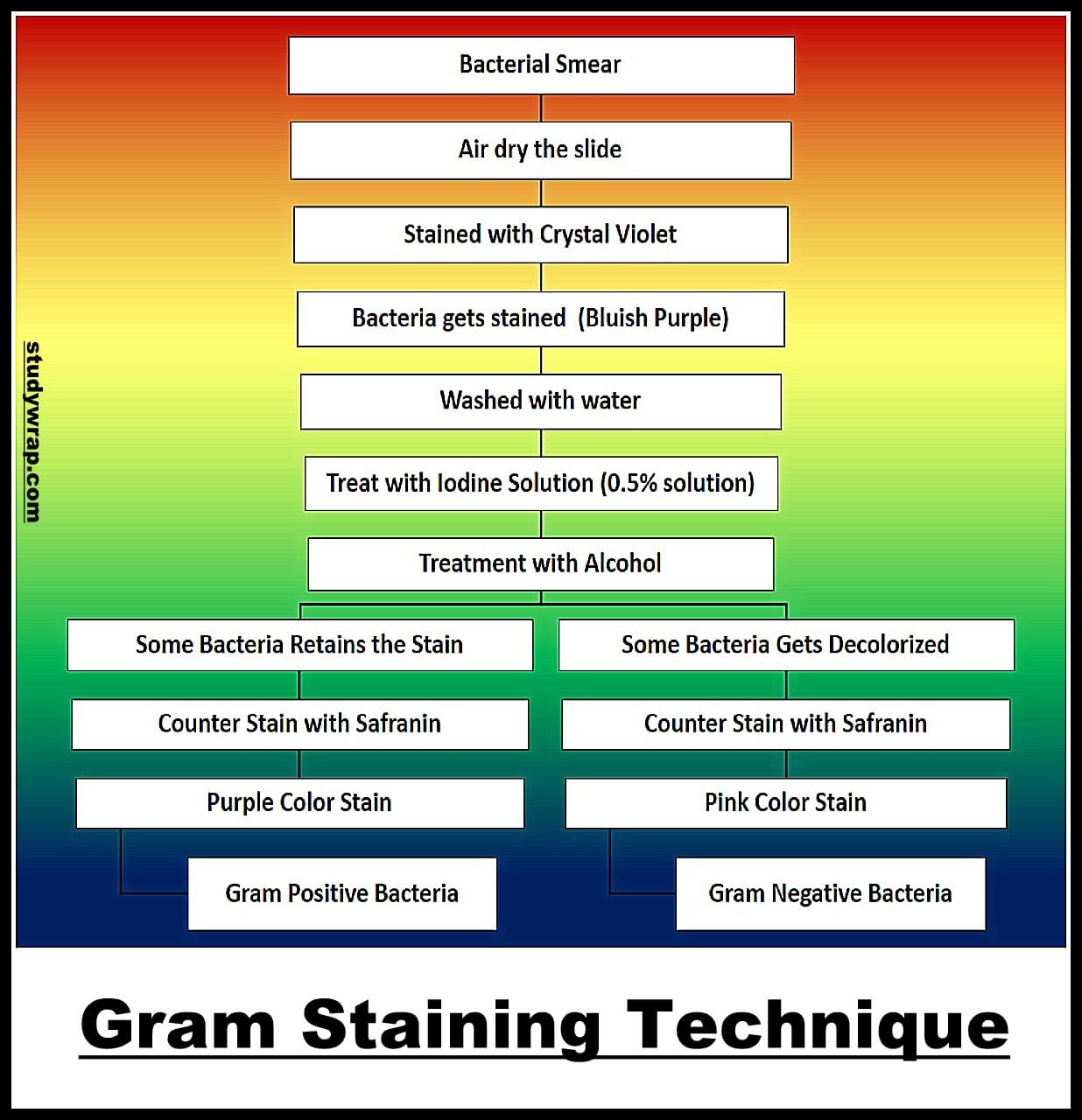
Procedure
· Prepare the Bacterial Slide.
· The bacterial smear is stained with weakly alkaline solution of crystal violet. Gently rinse off the stain with water.
· Iodine is subsequently added as a mordant to form the crystal violet-iodine complex, so that, the dye cannot be removed easily. This step is generally referred as fixing the dye.
· Treat it with alcohol decolorizer until the solution appears clear. Gently rinse with water.
· Cover the smear with safranin, the counterstain, for 20 seconds. Gently rinse the stain with water.
· A counterstain of safranin is applied to the smear to give decolorized gram negative bacteria a pink color.
· Gram positive bacteria appear violet due to the crystal violet stain and Gram negative bacteria which cannot retain the crystal violet stain, appear red or pink due to safranin.
Gram Positive Bacteria
· Gram-positive bacteria constitute a cell wall, which is mainly composed of multiple layers of peptidoglycan that forms a rigid and thick structure. Its cell wall additionally has teichoic acids and phosphate.
· In the cell wall of Gram Positive bacteria, both horizontal and vertical peptide linkages are present, due to which mesh is dense and hence the stain does not come out.
· The cell walls of Gram Positive bacteria contain 60-90% peptidoglycan.
· Cell wall of Gram Positive bacteria is thick (20-80 nm) and is destroyed by penicillin.
· Further outer layer of cell wall of Gram Positive bacteria is made of teichoic acid.
· Teichoic acid is an acidic polymer containing a carbohydrate (Eg. glucose), phosphate and an alcohol is found in cell walls of Gram positive bacteria.
· Teichoic acid has several roles to play in a cell like acting as receptor sites for some viruses, binding metals and maintaining cells pH in order to prevent its degradation by self-produced enzymes.
· The walls Gram positive bacteria contain very little amount of lipids.
· The cell wall of Gram Positive bacteria is more homogenous and single layered and amorphous.
Gram Negative Bacteria
· In gram-negative bacteria, the cell wall is made up of an outer membrane and several layers of peptidoglycan. The outer membrane is composed of lipoproteins, phospholipids, and lipopolysaccharides.
· The Gram negative bacteria have cell walls that are more complex than gram positive bacteria. The cell wall is thin (8-12 nm) and hard and is not destroyed by penicillin.
· In the cell wall of Gram Negative bacteria, either horizontal or vertical peptide linkages are present, due to which mesh is loose, hence, stain comes out. Further outermost layer of cell wall of Gram Negative bacteria is made of lipopolysaccharides.
· The peptidoglycan layer is very thin constituting only 10% or less of the total cell wall. There exists an outer membrane that covers the thin underlying layer of peptidoglycan.
· The outer membrane consists of phospholipid bilayer structure made chiefly of phospholipids, proteins and lipopolysaccharides (LPS).
· The outer membrane serves as a gate to preserve the important enzymes from leaving the periplasmic space between the cytoplasmic membrane and the outer membrane.
· It also monitors the entry of various chemicals that are harmful in nature to the cell.
· Further, cell wall of Gram Negative bacteria is less homogenous, more complex and multilayered. Lipids (including phospholipids, lipopolysaccharides and lipoproteins) make up 80-85% of the total cell wall.

Difference between Gram Positive Bacteria and Gram Negative Bacteria
|
Character |
Gram Positive |
Gram Negative |
|
Stain |
They take the Violet colour of Gram stain even after destaining with alcohol and appear as purple-coloured. |
They get the colour blue with Gram stain initially, however lose it after destaining with alcohol and appear as pink-coloured due to Safranin. |
|
Cell wall |
Single layered homogenous cell wall. |
Double layered heterogeneous cell wall. |
|
Thickness |
Thick (20-80 nm). |
Thin (8-12 nm). |
|
Cell Wall Content |
Cell wall is more rigid due to high percentage (80%) of peptidoglycan. |
Cell wall is less rigid due to low percentage (3-12%) of peptidoglycan. |
|
Muramic acid content is 70-95%. |
Muramic acid content is 5-20%. |
|
|
Lipid content is low (2-4%). |
Lipid content is high (20-30%). |
|
|
Phospholipid is absent in cell wall. |
Phospholipid is present in cell wall. |
|
|
Teichoic acid is present. |
Teichoic acid is absent. |
|
|
Resistance to Antibiotic |
More sensitive to antibiotics such as penicillin. |
Resistant to antibiotics like penicillin. |
|
|
Mostly cells are non-capsulated. |
Mostly cells are capsulated. |
|
Mesosome |
Mesosome are very common in the cell. |
Mesosome are rarely present in the cell. |
|
Pili |
Pili are usually absent. |
Pili are very common. |
|
Flagella Structure |
Flagella are less common. Basal body of flagellum has 2 rings (S, M) only. |
Flagella are very common. It has four rings (L, P, S and M). |
|
Toxin Produced |
Only few forms are pathogenic and may produce exotoxins. |
More forms are pathogenic and may produce endotoxins. |
|
Examples |
Bacillus, Clostridium, Lactobacillus, Streptococcus, Leuconostoc, Staphylococcus, Corynebacterium |
E coli, Salmonella, Acetobacter, Azotobacter, Vibrio, Agrobacterium, Shigella, Xanthomonas |
So, this was all about the Gram Positive and Gram Negative Bacteria and Gram Staining Technique. If you want to read more note related to Biology, then Click Here.
If you liked our Post, than do like our Facebook Page to get regular updates and share it with your Friends and Family. Sharing is Caring.
